-
Carestream DR Detector Family
• DRX Detectors
Retrofit Solutions
• DRX Mobile Retrofit
• Transportable Systems
Fuji Detector Family
• FDR D-EVO GL
• FDR D-EVO II
• FDR ES -
Carestream X-Ray Suite Family
• DRX-Evolution Plus
• DRX-Ascend
Fuji X-Ray Suite Family
• FDR D-EVO Suite II
• FDR Clinica -
Carestream Mobile X-Ray Family
• DRX-Revolution
• DRX-Revolution Nano
Fuji Mobile X-Ray Family
• FDR Go Plus
• FDR AQRO
-
• Esaote MyLab™ 9 Platform
• Esaote MyLab™ Eight Platform
• Esaote MyLab™ Twice
• Esaote MyLab™ ClassC
• Esaote MyLab™ Seven
• Esaote MyLab™ Six
• Esaote MyLab™ Alpha
• Esaote MyLab™ Gamma
• Esaote MyLab™ One
• Hitachi LISENDO 880
• Hitachi ARIETTA 70
• Hitachi ARIETTA 60
• Hitachi ProSound Alpha 7
• Hitachi Noblus
• Hitachi ProSound F37
• Hitachi ProSound F75
• Hitachi SOFIA
MyLab™ ClassC: Discover The Power Of Touch
Your Comfort With A Touch
Whenever physicians think of a high-level cardiovascular and general imaging ultrasound systems, they ask for up-to-date platforms, with high-performance and advanced on-board technologies as well as simplicity and ease of use.
MyLab™ClassC has been designed based on these key concepts to deliver a reliable diagnosis, and ensure every day productivity. With just one glance, you will understand how MyLab™ClassC‘s simplicity has never been seen on such a high-level ultrasound scanner.

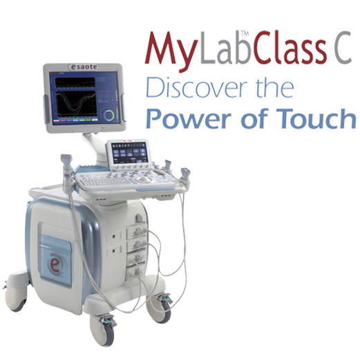
MyLab™ClassC: Performance, Simplicity And Ergonomics With A Single TouchHigh performance does not always mean large and stationary systems. A particular effort has been made to reduce size and increase the new MyLab™ClassC’s ergonomics. This has led to a compact and agile system, which is easy to move and is able to adapt to any kind of environment, including most critical ones such as interventional and operating rooms. The height-adjustable and rotating keyboard, as well as the multiplane-articulated monitor arm, allow for optimal positioning at all times. |
 |
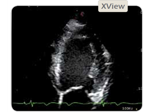
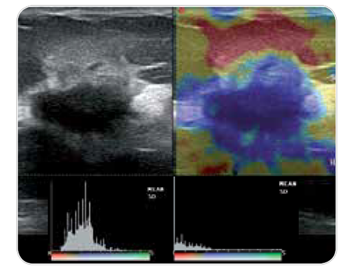
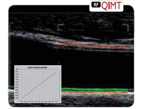
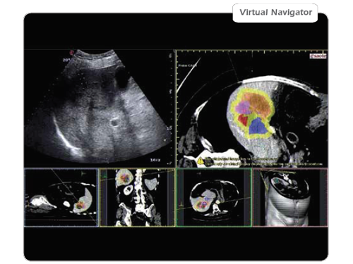
Advanced Technologies For Any Clinical SolutionMyLab™ClassC innovative characteristics allow the system to be placed in any environment, always providing easy, fast and reliable access to diagnosis and data sharing. Prevention and quantification in Cardiovascular Imaging are easily available with a touch: the prevention suite with RFQIMT, RFQAS, XStrain™, CFI advanced technologies and state-of-the-art iQProbes allows the best clinical approach to diagnoses. In General Imaging up-to-date CnTI™ Contrast Tuned Imaging for contrast media procedures, HD CFM and XFlow, High Frequency Image, X4D technology, Virtual Navigator and Elaxto-Elastosonography cover any clinical need. Imaging Processing: Esaote offers many technologies for imaging enhancement. With TEI™ the harmonic signal is fully preserved without degradation of the acoustic information. MView and XView improve quality of ultrasound images by reducing the presence of artifacts, shadowing and speckle.CnTI™ - Contrast Tuned Imaging for contrast media procedures: CnTI™, Esaote’s revolutionary technology, in combination with latest generation ultrasound contrast agents, provides impressive clinical results due to precise micro-bubble detection. The very low applied acoustic pressure, allows the bubbles life time to be increased, for a clear identification of arterial and late phase. The very high probe sensitivity and low level of noise and artifacts bring precise diagnosis, both for lesions detection and characterization. A contrast-dedicated quantification tool is also available. HD CFM and XFlow - Extraordinary flow sensitivity and spatial resolution: Color Doppler sensitivity and resolution are very important in the assessment of blood flow, especially for flows with limited dimensions and velocities. HD CFM technology helps the user define the right setting to obtain the maximum clinical information. In case of particular diagnostic processes in which morphological information are more important than the hemodynamic itself, XFlow delivers clear pictures with reduced artifacts, and less-important insonation angle dependence. High Frequency Image: Esaote's historical leadership in High-frequency Imaging delivers an unexpected level of details in any application in which superficial images are required. 22 MHz transducers, XView, MView, ElaXto and X4D, as well as "A Universe under the mm" package are just few examples of the technological potential of the MyLab™ClassC. The clinical results are simply astonishing, leading to new research fields and new levels of diagnosis. Advanced technologies such as ElaXto and X4D are implemented not just as additional qualitative information, but as important quantitative packages to deliver an objective and fast diagnosis. X4D Technology: The advanced 3D/4D package takes advantage of innovative ways of visualizing conventional 2D ultrasound images through sophisticated algorithms and is able to deliver outstanding 3D/4D volume reconstructions. Measurements of length, surface, perimeter, diameter and angle as well as volume areas in the multi-dimensional display, allow both quantitative analysis and qualitative acquisition. A link to a special database file includes all personal data sets. RFQIMT - RF-based Quality Intima Media Thickness for early detection of cardiovascular diseases: RFQIMT targets the measurements of blood vessel thickness in an area of the Carotid artery selected for investigation. Its ease of use, combined with real time quality feedback, helps the operator achieve accurate and reproducible results. The measurements (even when taken at different examination times) can be reported on a normalized graph displayed with plot indicators that will assist physicians in their diagnostic and therapeutic procedures. RFQAS - Quality Arterial Stiffness for early detection of cardio-vascular diseases: RFQAS targets the measurements of blood vessel stiffness in an area of the Carotid artery selected for investigation. The blood vessel’s wall stiffness is expressed in brachial blood pressure and accurate measurements of diameter and change in diameter. Local blood pressure at the site of the ultrasound measurement is also supplied. Local blood pressure and stiffness are derived as quantification results based on sophisticated clinical studies. Auto adjustment and measurement: Doppler profile quantification is an important issue in cardiology as well as vascular ultrasound examinations. Once the volume sample has been placed and the Doppler trace is displayed on the monitor, the user will be able to select the real-time assessment of all key clinical parameters by enabling the ADM function. When working with freeze-frame mode is preferred, you can still trace Doppler contour and track maximum, mean or minimum values automatically. Features like EF Calculation and ADM (automatic measurement) provide quantification of important clinical parameters in a short time. This allows for faster screening and accurate patient management in case of potential diseases that may be further investigated. CFI - Coronary Flow Imaging to assess coronary artery blood flow and its main characteristics: The assessment of Coronary Blood Flow characteristics is meaningful in alnalyzing basal cardiac activity without any externally induced cardiac stress. When the CFI Colour Doppler preset is enabled, the signal coming from the coronary artery blood flow is optimized against many concomitant velocity components of blood flow present within the heart ventricles and atria. The combination of a Cardiac iQ-probe and the dedicated CFI (Coronary Flow Imaging) preset offers a superior performance in CFM/PW modes for the detection and measurement of Coronary Flows. XStrain™: XStrain™ is a non-invasive tool for an enhanced investigation of myocardial function, allowing physicians to explore and quantify aspects of the heart’s physiology, which were not detectable or quantifiable with previous ultrasound technologies. Myocardial velocity, myocardial strain and strain rate can detect early impiarment of pump functions (assessed as ejection fraction or stroke volume). Since it relies on angle-independent technology, XStrain™ allows assessment of both right and left ventricle contractibility. XStrain™ provides an innovative tool for the mechanical assessment of the heart’s wall motion. It can therefore provide quantitative support for standard echo examinations and be used to examine and monitor patients to identify cardiac wall motion early change signs. Virtual Navigator advanced tool for Fusion Imaging (US & CT/MR/PET-CT): Virtual Navigator, with Fusion Imaging applied to US enhances the information produced by an Ultrasound Scanner by combining images with a second modality (CT, MR, PET or 3D US) in real-time: yielding all the benefits of different modalities in the same exam. Virtual Biopsy - Advanced Biopsy also in very difficult approaches: Virtual Biopsy allows physicians to follow percutaneous procedures by superimposing the needle tracking information on the real-time ultrasound image. |
|
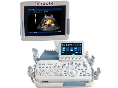
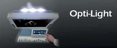
Opti-Light: High-Performance Optimized LightingOptimal lighting has always been a crucial factor for ultrasound imaging. The latest LCD Monitor Technology allows images to be clearly displayed under any condition. MyLab™ClassC also introduces an additional unique feature: Opti-Light. This feature, thanks to a light point behind the monitor, allows the operator to control the room's lighting level directly from the system, through the especially designed controls located on the touch screen. Optimized working conditions increase users’ comfort and improve patient care. |
|

|
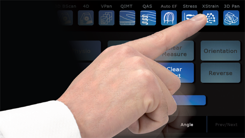
High-Quality Touch Screen: Completeness With A TouchThe large high-quality touch screen is well positioned near the most important working area of the control panel. This touch-screen allows all mode-dependent parameters to be clearly displayed and changed with one simple touch. |
|
Contact Prestige Medical Imaging Today To Address All Of Your X-Ray Imaging Questions